|
A few months ago I wrote here about my work using X-Ray micro-CT to study the coevolution of male and female genitalia in insects. I also used that post to present some early images of the male and female genitalia of the seed beetle Callosobruchus maculatus I made from those scans. In this post I would like to present some more CT images, but focusing on a different species: the Indian Meal Moth Plodia interpunctella. This time we are looking at the interaction between the region of the female reproductive tract where the male deposits his sperm (known as the bursa), and the structure containing the male’s sperm, known as the spermatophore. In several insect groups, including butterflies and moths, the male sperm is not deposited freely but is instead transferred in a discrete package known as a spermatophore. In Plodia interpunctella the spermatophore has an elaborate structure. Below is a 3d volume rendering of a spermatophore inside a female. The spermatophore has two main parts: a hollow, spherical sac which contains the sperm mass, and an elaborate chitinous neck. 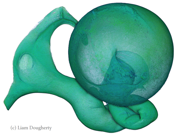 3d reconstruction (false-colour) of a male spermatophore inside the female bursa after mating. Note the spherical sac (right) and long neck region ending with a hole (left). The spermatophore is passed to the female in liquid form, which then solidifies and hardens during mating into the characteristic shape seen here. Inside the spherical sac are around 30 large sperm bundles, each formed of many sperm cells (see below). Once in the spermatophore has been deposited, the sperm move along the neck and out of the hole at the top. 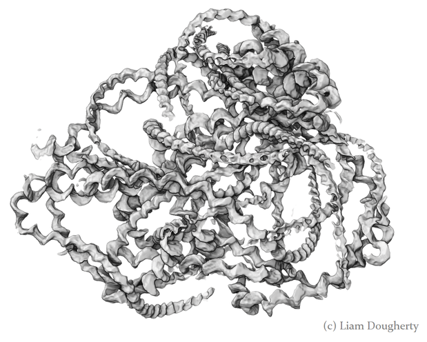 3d reconstruction of the sperm bundles tightly packed inside the spermatophore. Once the sperm has been transferred to the female the spermatophore shrinks but is not broken down by the female (see below). This is useful because it means that we can count the number of times a female has mated (either in the wild or in the lab) by dissecting out the bursa after she has died and counting the number of spermatophores present. 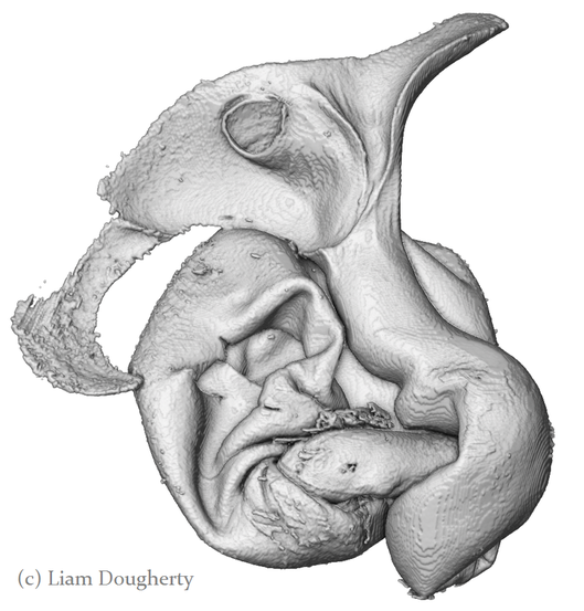 3d reconstruction of an old, shrunken spermatophore inside the bursa of a female. These can be counted to determine the number of times a female has mated. The female bursa is a large hollow organ into which the spermatophore is deposited. Sperm are not stored here, but instead are stored in a much smaller organ called the spermatheca. To get to the spermatheca the sperm need to be moved along a narrow duct which opens into the bursa. Surrounding the entrance to this duct on the wall of the bursa is a row or sharp teeth known as lamina dentata or signa (see below). The teeth vary in size and shape within this species. 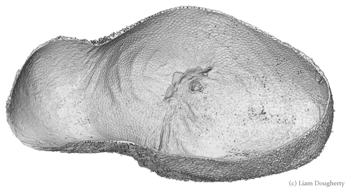 3d reconstruction of the female bursa, with a virtual slice to show the internal structure. Note the row of teeth next to the spermathecal duct entrance. In other moth and butterfly species these teeth are used to physically pierce the spermatophore capsule, thus releasing the spermatophore contents into the bursa. One reason why females may want to pierce the spermatophore is because the presence of a full spermatophore in the bursa may prevent the female from remating. Males generally benefit from preventing female remating as it may reduce his paternity share. Therefore one hypothesis fur the function of bursal teeth is that they allow the female to regain some control of her remating rate by speeding up the breakdown of the spermatophore. Males could then evolve larger or thicker spermatophores in response, leading to an ‘arms race’ between bursal anatomy and spermatophore anatomy. I have been using micro-CT to examine potential coevolution between bursal teeth morphology and spermatophore morphology in the Indian Meal Moth. This work is being done in collaboration with Leigh Simmons at Kathryn McNamara at UWA, and Nina Wedell at the University of Exeter. Watch this space for news on future publications!
0 Comments
|
Archives
February 2024
AuthorLiam Dougherty. Categories |
 RSS Feed
